Anatomy Of Musckes Sndctendons / Anatomy Of Knee
• definitions • introduction • development of muscles • classification • anatomy of skeletal muscle • muscle physiology • properties • muscles of development of muscles. Smooth muscles are found in the walls of many organs, such as the stomach and in blood vessels. Anatomy of the short head of the biceps brachii muscle. Skeletal muscles are attached to bones by tendons and can be as long as 30 cm, although they are usually 2 to 3 cm in length. Learn about the muscles, tendons, bones, and ligaments that comprise the knee joint anatomy. It occupies most of the oral cavity and oropharynx. An interactive tutorial teaching the position, actions, innervation and attachments of the rectus femoris muscle with the aid of anatomical illustrations. The muscular system is composed of specialized cells called muscle fibers.
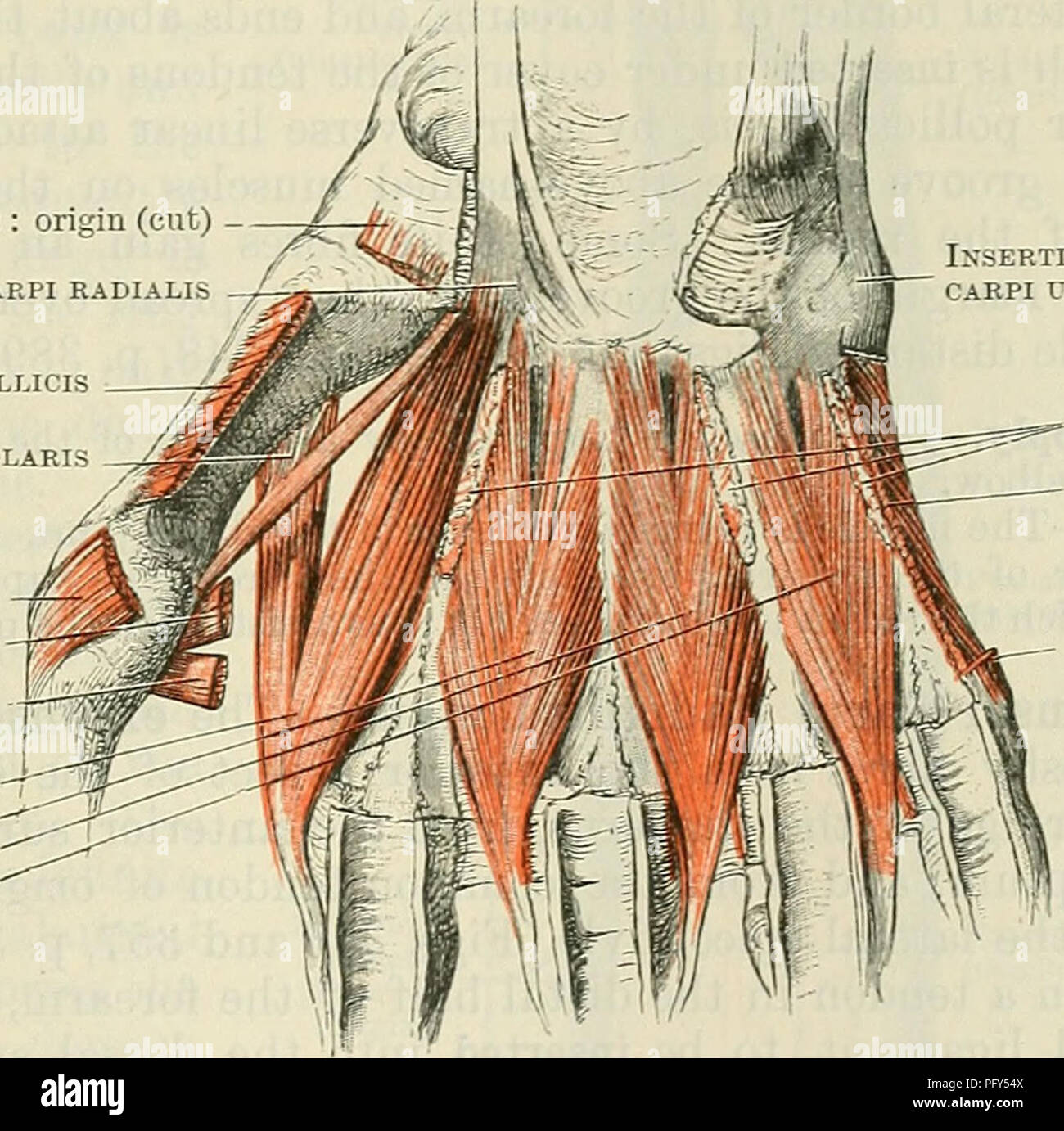
Anatomy of the short head of the biceps brachii muscle. Find the best weight lifting exercises that target each muscle or groups of muscles. The tendons of these muscles pass through a small corridor in the wrist known as the carpal tunnel.
The skeletal muscles are continually making fine adjustments that hold the body in stationary positions.
Learn about human anatomy muscles with free interactive flashcards. Along with lateral pterygoid muscle it produces side to side movement of mandible. This is a table of skeletal muscles of the human anatomy. • muscle tissues develop from embryonic cells. The skeletal muscles are continually making fine adjustments that hold the body in stationary positions. Cardiac muscle contracts the heart to pump blood. In the muscular system, muscle tissue is categorized into three distinct types: Muscular contraction is necessary for voluntary and involuntary movement of limbs, stabilization of joints, maintaining luminal diameter (in the case of arteries, bowel, etc), and to produce heat. Anatomy of the short head of the biceps brachii muscle. Each of these muscles is a discrete organ constructed of skeletal muscle tissue, blood vessels, tendons, and nerves. Discover the muscle anatomy of every muscle group in the human body. Circular skeletal muscles are made up of fibers that are arranged in a circular manner. Skeletal muscle is a voluntary muscle, which means that we can actively control its function. In this section, learn more about the anatomy of the muscles of the neck. There are two main muscle groups around the knee:
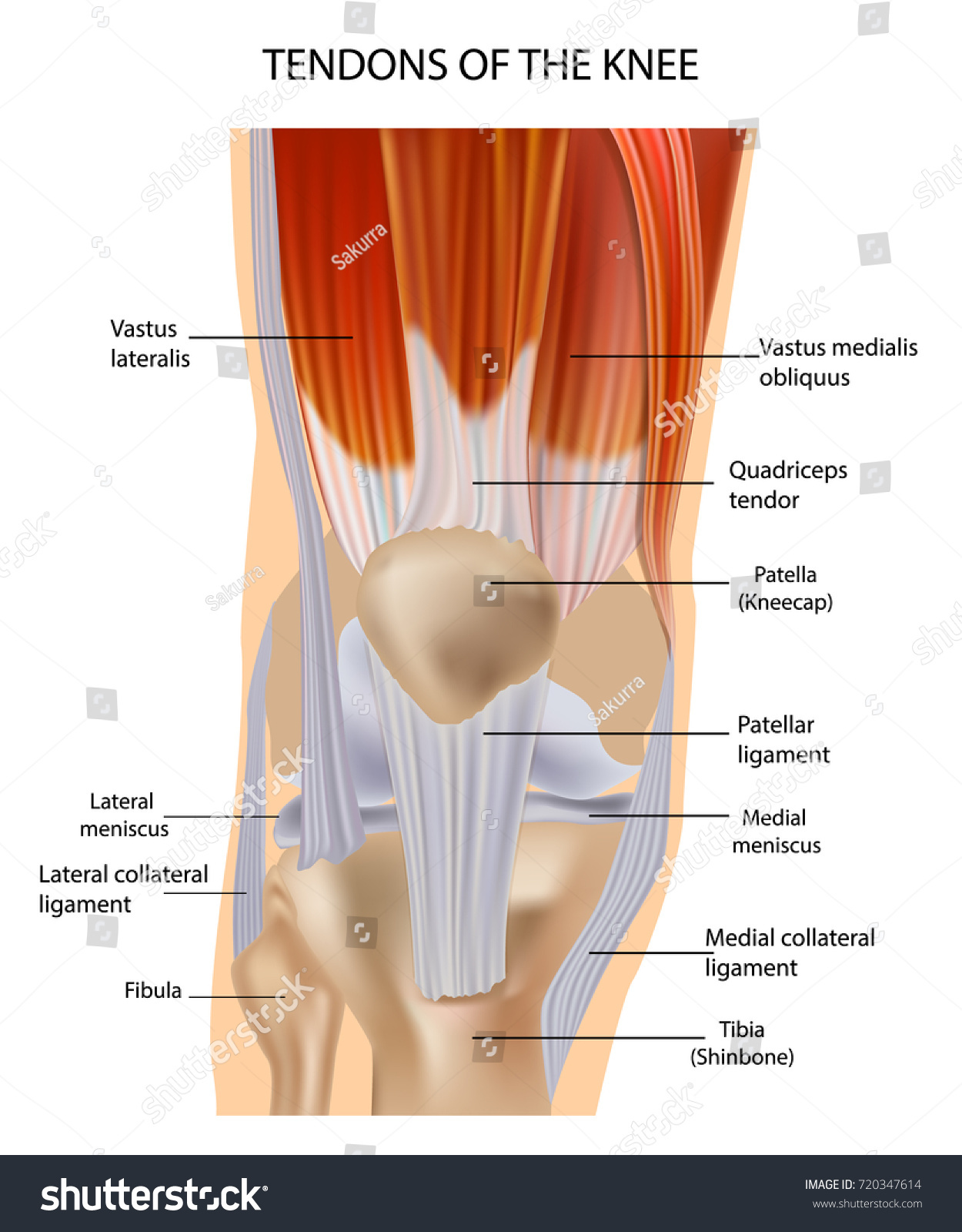
Knee function is determined in large part by the anatomy of the joint. The muscular system consists of about 700 muscle organs that are typically attached to bones across a joint to produce all voluntary movements. The muscular system is responsible for the movement of the human body. Inflammation of this region caused by repetitive stress or trauma may lead to pain and numbness known as carpal tunnel syndrome. See the pictures and anatomy description of knee joint bones, cartilage, ligaments, muscle and tendons with resources for knee problems & injuries. The muscles around the knee help to keep the knee stable, well aligned, and moving. Convergent muscles contain fibers that have a wide origin, but converge in order to attach to a narrow tendon. Muscular contraction is necessary for voluntary and involuntary movement of limbs, stabilization of joints, maintaining luminal diameter (in the case of arteries, bowel, etc), and to produce heat. The muscular system is composed of specialized cells called muscle fibers.

Cardiac muscle contracts the heart to pump blood.
Inflammation of this region caused by repetitive stress or trauma may lead to pain and numbness known as carpal tunnel syndrome. There are two main muscle groups around the knee: • muscle tissues develop from embryonic cells. • definitions • introduction • development of muscles • classification • anatomy of skeletal muscle • muscle physiology • properties • muscles of development of muscles. The tendons of many muscles extend over joints and in this way. The muscular system is composed of specialized cells called muscle fibers. The inguinal aponeurotic falx (falx aponeurotica inguinalis; You can click the links in the image, or the links below the image to find out more information on any muscle group. An interactive tutorial teaching the position, actions, innervation and attachments of the rectus femoris muscle with the aid of anatomical illustrations. You can't control them, but smooth muscles are at work all over your body. General functions of muscular system: There's no strict demarcation or dividing line between the tendon and the covering around this muscle but that covering is called is called the epimysium fp my cm and it's really just connective tissue that covers the muscle kind of protects it reduces friction. Cardiac muscle contracts the heart to pump blood. Muscular contraction is necessary for voluntary and involuntary movement of limbs, stabilization of joints, maintaining luminal diameter (in the case of arteries, bowel, etc), and to produce heat.
Practice identifying the major muscles of the human body. In the muscular system, muscle tissue is categorized into three distinct types: The tendons of many muscles extend over joints and in this way. Conjoined tendon of internal oblique and transversalis muscle) of the obliquus internus and transversus is mainly. Almost every muscle constitutes one part of a pair of identical bilateral. From anterior to posterior, the tongue has 3 surfaces:
The tip is the highly mobile, pointed anterior portion of the tongue.
Cardiac muscle contracts the heart to pump blood. The muscles around the knee help to keep the knee stable, well aligned, and moving. It elevates and protrudes the mandible. There are two main muscle groups around the knee: The tongue is a mass of muscle that is almost completely covered by a mucous membrane. It occupies most of the oral cavity and oropharynx. A neat little trick to learn the superficial muscles of the forearm is to use your fingers as the guide. Each of these muscles is a discrete organ constructed of skeletal muscle tissue, blood vessels, tendons, and nerves. The muscular system is responsible for the movement of the human body. Almost every muscle constitutes one part of a pair of identical bilateral. This is a table of skeletal muscles of the human anatomy. The skeletal muscles are continually making fine adjustments that hold the body in stationary positions. Convergent muscles contain fibers that have a wide origin, but converge in order to attach to a narrow tendon. The anatomy of muscle cells differs from that of other body cells and biologists have applied specific terminology to different parts of these cells. Anatomy of the short head of the biceps brachii muscle.

The smooth muscle tissue that forms organs like the stomach and bladder changes.

Attached to the bones of the skeletal system are about 700 named muscles that make up roughly half.

• the muscular system develops from intra embryonic mesoderm.

Anatomy of the short head of the biceps brachii muscle.

• muscle tissues develop from embryonic cells.

The three scalene muscles are found forming the floor of the posterior triangle.

Circular skeletal muscles are made up of fibers that are arranged in a circular manner.

The three scalene muscles are found forming the floor of the posterior triangle.

From anterior to posterior, the tongue has 3 surfaces:

The tendons of these muscles pass through a small corridor in the wrist known as the carpal tunnel.

Cardiac muscle contracts the heart to pump blood.

General functions of muscular system:

The tendons of these muscles pass through a small corridor in the wrist known as the carpal tunnel.

Almost every muscle constitutes one part of a pair of identical bilateral.

• the muscular system develops from intra embryonic mesoderm.

An interactive tutorial teaching the position, actions, innervation and attachments of the rectus femoris muscle with the aid of anatomical illustrations.

Learn about human anatomy muscles with free interactive flashcards.

The muscular system is responsible for the movement of the human body.

Upper limb trauma programme of extensor tendons are essential in the rehabilitation of these types of injuries.

It occupies most of the oral cavity and oropharynx.

The anatomy of muscle cells differs from that of other body cells and biologists have applied specific terminology to different parts of these cells.

Knee function is determined in large part by the anatomy of the joint.

A neat little trick to learn the superficial muscles of the forearm is to use your fingers as the guide.

Attached to the bones of the skeletal system are about 700 named muscles that make up roughly half.
Find the best weight lifting exercises that target each muscle or groups of muscles.

Along with lateral pterygoid muscle it produces side to side movement of mandible.
 are usually in sheets or layers, with one layer of muscle behind the other.")
Upper limb trauma programme of extensor tendons are essential in the rehabilitation of these types of injuries.

All 4 muscles have a common origin at the medial epicondyle of the humerus, known as the common flexor tendon.

In the diagrams below, i'll be showing muscle groups in color, with a black line to show the forms that would show through the skin (i also show protruding bones that would do the same).

Skeletal muscle is a voluntary muscle, which means that we can actively control its function.

Their predominant function is contractibility.

The tendons of these muscles pass through a small corridor in the wrist known as the carpal tunnel.

Cardiac muscle contracts the heart to pump blood.

The tendons of these muscles pass through a small corridor in the wrist known as the carpal tunnel.

The anterior and middle scalenes originate from the transverse processes of certain cervical vertebrae and attach to the first rib.

Posting Komentar untuk "Anatomy Of Musckes Sndctendons / Anatomy Of Knee"